
Thyroid Disease
HOW DOES THE THYROID GLAND FUNCTION?
- The thyroid gland located in the neck has the primary function of secreting a hormone called thyroxine. As this hormone contains 4 iodine atoms, it is also referred to as T4.
- T4 is an inactive molecule that requires activation to tri-iodothyronine (or T3). This process requires removal of an iodine atom from T4.
- The thyroid gland is controlled by another gland that is located in the brain called the pituitary gland. This gland releases a hormone called thyroid stimulating hormone, or TSH. As the name suggests, TSH is responsible for stimulating the thyroid gland to release T4.
- The release of T4 from the thyroid gland is dependent on the quantity of TSH released by the pituitary. Similarly, the quantity of TSH released by the pituitary gland is dependent on the amount of T4 in the blood stream. This mechanism of control is sometimes called a ‘feedback’ mechanism.
- The process is very simple. When the pituitary gland secretes TSH, it travels through the blood stream to the thyroid gland. The TSH then dictates the release of T4 from the thyroid gland. Once sufficient T4 has been released, the pituitary gland recognizes this and stops producing TSH.

In other words, T4 levels dictate how much TSH will be released.
- When the thyroid gland is underactive (hypothyroidism), the TSH is unable to stimulate release of T4 from the thyroid gland. As a result, the pituitary attempts to push out more TSH, but this has no effect. This is why patients with hypothyroidism have elevated TSH levels and low T4 levels.
- The opposite is true when the thyroid is overactive (hyperthyroidism). The T4 levels will be markedly elevated, while the TSH levels could be undetectable on blood tests.
- Once the thyroid hormones T3 and T4 have been released, they are transported through the blood stream with the help of transport proteins. This would mean that if the protein levels were low, the quantity of thyroid hormone reaching vital tissues would be less. The opposite is true as well.
Changes in thyroid transport proteins levels can be seen in pregnancy and in women taking the oral contraceptive pill.
If there is some concern about the protein levels, then a test called free T4 (FT4) test could be done. This measures the level of thyroid hormone that is unbound to protein.
Testing Thyroid Function
Thyroid function is easily evaluated through blood tests. Below are the commonly performed tests to asses thyroid function.
TSH Levels
-
-
- As previously discussed, TSH levels are elevated in hypothyroidism and reduced in hyperthyroidism. The levels of TSH can be checked with a simple blood test.
- Low TSH levels can be due to 2 reasons. The first reason is elevated T4 levels from an overactive thyroid gland. The second reason is a malfunctioning pituitary gland, which itself does not release TSH.
- When performing a routine thyroid evaluation, it is not always necessary to check all the parameters. A TSH test is often sufficient, as if this is normal, it is likely the remainder of the function is normal as well. However, it is important to interpret the results based on clinical history.
-
T4 levels
-
-
- T4 exists in 2 forms in the blood stream – a protein bound form and an unbound form.
- T4 that is bound to protein does not allow the T4 to enter vital structures to exert it effects.
- However, unbound T4, or free T4 is the one that is more important, as this reflects what proportion of the thyroid hormone is exerting its effects on the vital structures.
- Previously, a free thyroxine index (o FTI) was performed to assess the function of the thyroid gland. After the advent of free T4 tests, this test is now rarely performed. This is also because FTI levels can sometimes be elevated even when the T4 is normal, thus giving a false diagnosis of hyperthyroidism.
- If a thyroid function test reveals low FT4 levels and increased TSH levels, it indicates the presence of a condition called primary hypothyroidism.
- If the TSH is low and the FT4 is also low, it indicates a problem with the pituitary gland called secondary hypothyroidism.
- Finally if the TSH is low and the FT4 is high, it is indicative of hyperthyroidism.
-
T3 Levels
-
-
- The main reason for checking T3 levels is to make a diagnosis of an overactive thyroid, or hyperthyroidism. Elevated levels of T3 are seen in hyperthyroidism.
- On the other hand, T3 levels are rarely useful when it comes to diagnosing hypothyroidism. This is because the levels take quite a while to reduce, despite the T4 levels being low.
- AS explained previously, altered levels of T3 can also be seen when protein levels are abnormal.
- Thyroid antibody tests
- Antibodies are the body’s defence against infections. They form a part of our immune system and are produced by cells called lymphocytes.
- Sometimes, antibodies may be produced by the body against the thyroid gland. This can lead to an over- or underactive thyroid gland as they may either stimulate it or suppress it respectively.
- There are 2 main types of antibodies that are directed against thyroid proteins that can affect the thyroid gland – anti- thyroid peroxidise antibodies ( anti-TPO) or anti-thyroglobulin. Sometimes, tests may need to be conducted to determine their levels in the blood.
- For example, in a patient with hypothyroidism and anti-TPO/anti-thyroglobulin antibodies is likely suffering from a condition called Hashimoto’s thyroiditis.
- Similarly, in autoimmune thyroid disease, these antibodies lead to hyperthyroidism.
-
Thyroglobulin
-
-
- The presence of thyroglobulin protein is indicative of normal thyroid function or thyroid cancer. IN other words, the presence of thyroglobulin is not diagnostic of thyroid cancer.
- However, in patients who have undergone surgery and had their thyroid gland removed due to thyroid cancer, increasing levels of thyroglobulin could indicate recurrence.
-
Other Tests
Radioactive Iodine Uptake
-
-
- The thyroid gland requires iodine to function. This uptake of iodine is reflective of its function. If the thyroid gland is functioning normally, the quantity of iodine taken up should be within a particular range.
- Radioactive iodine is sometimes used when investigating thyroid disease.
- By using radioactive iodine, it is possible to determine how much iodine the thyroid gland pulls in. If excessive amount of radioactive iodine is taken up by the gland, it indicates an overactive thyroid gland. If very little is taken up, it is indicative of hypothyroidism.
- Radiological studies may also help diagnose thyroid disease.
-
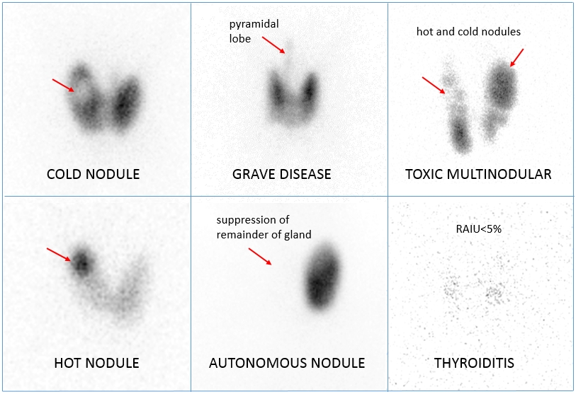
What is a Thyroid Scan and Uptake?
-
-
- A thyroid scan is a type of nuclear medicine imaging. The radioactive iodine uptake test (RAIU) is may also be called a thyroid uptake. It is a measurement of thyroid function, but does not involve imaging.
- Nuclear medicine is a branch of medical imaging that uses small amounts of radioactive material to diagnose and determine the severity of or treat a variety of diseases, including numerous types of cancers, heart disease, gastrointestinal, endocrine, neurological disorders and other abnormalities within the body. Because nuclear medicine procedures are able to pinpoint molecular activity within the body, they offer the potential to identify disease in its earliest stages as well as a patient’s immediate response to therapeutic interventions.
- Nuclear medicine imaging procedures are noninvasive and, with the exception of intravenous injections, are usually painless medical tests that help physicians diagnose and evaluate medical conditions. These imaging scans use radioactive materials called radiopharmaceuticals or radiotracers.
- Depending on the type of nuclear medicine exam, the radiotracer is either injected into the body, swallowed or inhaled as a gas and eventually accumulates in the organ or area of the body being examined. Radioactive emissions from the radiotracer are detected by a special camera or imaging device that produces pictures and provides molecular information.
- The thyroid scan and thyroid uptake provide information about the structure and function of the thyroid. The thyroid is a gland in the neck that controls metabolism, a chemical process that regulates the rate at which the body converts food to energy.
-
When something goes wrong.
If the gland produces too much (hyper) hormone we term the condition Hyperthyroidism. If the gland makes to little (hypo), we call this Hypothyroidism.
Hyperthyroidism or Graves' disease is an autoimmune disease.
-
-
- It most commonly affects the thyroid, causing it to grow to twice its size or more (goiter), be overactive, with related hyperthyroid symptoms such as increased heartbeat, muscle weakness, disturbed sleep, and irritability.
- It can also affect the eyes, causing bulging eyes (exophthalmos). It affects other systems of the body, including the skin and reproductive organs.
- It affects up to 2% of the female population, often appears after childbirth, and has a female:male incidence of 5:1 to 10:1.
- It has a strong hereditary component; when one identical twin has Graves' disease, the other twin will have it 25% of the time. Smoking and exposure to second-hand smoke is associated with the eye manifestations but not the thyroid manifestations.
- Diagnosis is usually made on the basis of symptoms, although thyroid hormone tests might possibly be useful, particularly to monitor treatment
-
Symptoms
-
-
-
- fast heart rate
- eyes might possibly bulge forward
- nervousness
- increased sweating
- muscle weakness
- trembling hands
- weight loss
- skin changes
- increased frequency of bowel movements
- decreased menstrual flow and less frequent menstrual flow
-
-
Hypothyroidism
-
-
- occurs when there is too little thyroid hormone in the blood
- affects more than 5 million people
- 10 times more common in women than in men
- one out of every 4,000 infants is born hypothyroid
- Symptoms
- feeling slow or tired
- drowsiness
- poor memory
- muscle cramps
- dry and course skin
- milky discharge from the brests
- husky voice
- feeling cold
- slow heart rate
- difficulty concentrating
- weight gain
- heavy menstrual flow
- infertility
- feeling depressed
-
- When thyroid disease affects the eyes, it is called Thyroid Eye Disease ({formely} known as Graves' ophthalmopathy). Eyes might possibly bulge or appear red and swollen.
- The space between the lids (palpebral fissure) might possibly widen. Excess tearing and discomfort might possibly occur in either or both eyes (see below).
- Patients might possibly experience sensitivity to light, blurring or double vision, inflammation, or decreased movement.
Procedures

